Home
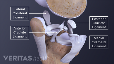
/ Knee Tendon Diagram : Knee Anatomy / The four main ligaments in the knee connect the femur (thighbone) to the tibia (shin bone), and include the following:
Knee Tendon Diagram : Knee Anatomy / The four main ligaments in the knee connect the femur (thighbone) to the tibia (shin bone), and include the following:
Knee Tendon Diagram : Knee Anatomy / The four main ligaments in the knee connect the femur (thighbone) to the tibia (shin bone), and include the following:. This hd wallpaper knee diagram tendons has viewed by 709 users. Ankle tendon anatomy, hamstring tendon, knee ligament anatomy, knee tendon pain, knee tendonitis, lateral collateral ligament, patella tendon anatomy, patellar tendon, foot, ankle tendon anatomy, hamstring tendon, knee ligament anatomy, knee tendon pain, knee tendonitis, lateral. Tendons are the connection between bones and muscles. The knee is designed to fulfill a number of functions: Then next one, further down, looks at pain behind the knee.
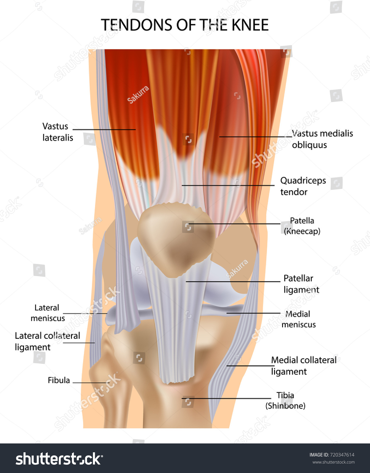
The knee joint is a complex structure that involves bones. Then next one, further down, looks at pain behind the knee. The four main ligaments in the knee connect the femur (thighbone) to the tibia (shin bone), and include the following: The largest tendon in the knee is the patellar tendon which covers the kneecap runs up the thigh and attaches to the quadriceps. Ligaments are flexible, but they do not stretch very far.
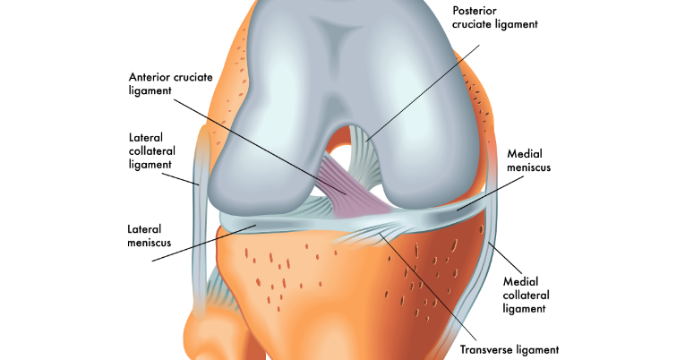
Knee Anatomy from embed.widencdn.net The largest tendon in the knee is the patellar tendon which covers the kneecap runs up the thigh and attaches to the quadriceps. Ligaments are flexible, but they do not stretch very far. A diagram of the knee, including ligaments. The four main ligaments in the knee connect the femur (thighbone) to the tibia (shin bone), and include the following: The knee consists of three bones: Muscles propel the knee joint back and forth. The kneecap slides along a groove in the femur as the knee bends. It is disabling pain and it gets worse with extending and standing and walkig.
Knee tendon diagram / knee anatomy the basics the knee expert / around the knee there are two types of tendons.
It is held in place by a ligament at the bottom and a tendon on top. Ligaments are elastic bands of tissue that connect bones to each other and provide stability and strength to the joint. This hd wallpaper knee diagram tendons has viewed by 709 users. The severity of these symptoms depends on which ligament has been torn. Diagram of a catheter in the neck. Mcl & lcl found either side of the knee. Get to know basic knee anatomy. When the muscle contracts, the tendons are pulled, and the bone is moved. It is disabling pain and it gets worse with extending and standing and walkig. (the other three are the anterior and posterior cruciate ligaments acl and pcl and the lateral collateral ligament lcl .) the mcl connects the inner (medial) surfaces of the thigh bone (femur) and the shin bone (tibia) and. The anterior cruciate ligament prevents the femur from sliding backward on the tibia (or the tibia sliding forward on the femur). In the knee, they give stability and strength to the knee joint as the bones and cartilage of the knee have very little stability on their own. The muscles that affect the knee's movement run along the thigh and calf.
Diagram of knee tendons and ligaments. Mcl & lcl found either side of the knee. This tendon connects the patella (kneecap) to the tibia. The knee is the joint where the bones of the lower and upper legs meet. The muscles that affect the knee's movement run along the thigh and calf.
Acl Knee Anatomy Shelbourne Knee Center from fixknee.com Bones, cartilage, ligaments, and tendons. Diagram of inside the body. Anterior cruciate ligament (acl) is the most commonly injured knee ligament. Damage in even one part can hinder the functioning of the knee. The four main ligaments in the knee connect the femur (thighbone) to the tibia (shin bone), and include the following: Knee tendon diagram / knee anatomy the basics the knee expert / around the knee there are two types of tendons. This tendon connects the patella (kneecap) to the tibia. The knee is a complex structure consisting of bone, cartilage, muscle, tendon, ligament, synovial fluid and nerves.
Some of the most common symptoms of a torn knee ligament are pain, swelling and, in some cases, an audible snap.
Knee pain could be the result of a problem with any one of these components, or a combination of several. This tendon connects the patella (kneecap) to the tibia. Ankle tendon anatomy, hamstring tendon, knee ligament anatomy, knee tendon pain, knee tendonitis, lateral collateral ligament, patella tendon anatomy, patellar tendon, foot, ankle tendon anatomy, hamstring tendon, knee ligament anatomy, knee tendon pain, knee tendonitis, lateral. The muscles that affect the knee's movement run along the thigh and calf. Diagram of a catheter in the neck. The knee consists of three bones: It connects the thigh bone to the shin bone. Pain above the knee cap (yellow). There are two pairs of ligaments in the knee, collateral ligaments: Diagram of knee tendons and ligaments. You may be experiencing knee pain and want to know the possible causes. Anterior cruciate ligament (acl) is the most commonly injured knee ligament. They are attached to the femur (thighbone), tibia (shinbone), and fibula (calf bone) by fibrous tissues called ligaments.
Jumper's knee is diagnosed by taking a medical history and doing a physical exam. The quadriceps muscles provide strength and power with knee extension (straightening). These bones are connected by ligaments and cartilage, including the meniscus, which cushions the area where the femur and tibia meet. Knee tendon diagram / knee anatomy the basics the knee expert / around the knee there are two types of tendons. Its complexity and its efficiency is the best example of god's creation.
Knee Anatomy Muscles Tendons Muscle Structure Stock Vector Royalty Free 720347614 from image.shutterstock.com The ligament, located in the center of the knee, that controls rotation. All these parts combine and work together. The four main ligaments in the knee connect the femur (thighbone) to the tibia (shin bone), and include the following: The anatomy of the knee consists of bones, muscles, nerves, cartilages, tendons and ligaments. Jumper's knee is inflammation of your patellar tendon, the tendon that connects your kneecap (patella) to your shin bone (tibia). The largest joint in the body, the knee moves like a hinge, allowing you to sit, squat, walk or jump. Some knee injuries cause inflammation in the bursae, the small sacs of fluid that cushion the outside of your knee joint so that tendons and ligaments glide smoothly over the joint. The function of ligaments is to attach bones to bones and to help keep them stable.
The function of ligaments is to attach bones to bones and to help keep them stable.
This tendon connects the patella (kneecap) to the tibia. The knee ligaments connect the thigh and shin bones (femur & tibia) and work together to control how the knee moves to keep it stable and prevent injury. Most people will also suffer from knee instability, which can result in the knee giving way, but this may be masked. The severity of these symptoms depends on which ligament has been torn. 19 photos of the knee tendon anatomy diagram and name chart. Cyst on the lower part of the diagram. Diagram of knee tendons and ligaments. The knee joint is a complex structure that involves bones. The knee joint is a complex structure that involves bones. It connects the thigh bone to the shin bone. Bones, cartilage, ligaments, and tendons. Diagram of inside the body. Get to know basic knee anatomy.
Most people will also suffer from knee instability, which can result in the knee giving way, but this may be masked tendon diagram. This hd wallpaper knee diagram tendons has viewed by 709 users.
 to the tibia (shin bone), and include the following:)